
Cavum velum interpositum Image
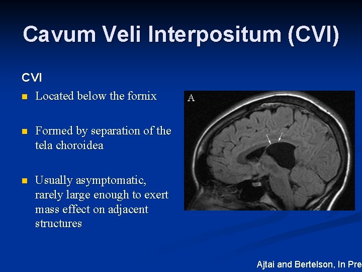
The velum interpositum (VI) is a membrane resulting from the superposition of two layers of the tela choroidea of the third ventricle demarcating a potential space containing cerebrospinal fluid (CSF) located in the region between the internal cerebral veins (ICV) and the posterior medial choroidal artery.

Cavum velum interpositum cyst Image
The internal cerebral veins are situated between the two layers of the cavum velum interpositum as is the posterior medial choroidal artery . On axial imaging, the cavum velum interpositum has a distinct triangular appearance with an apex directed anteriorly toward the forniceal columns (Fig. 1). An enlarged cavum velum interpositum displaces.

Cavum velum interpositum cyst Radiology Case
A cavum veli interpositi (CVI), often incorrectly termed a cavum velum interpositum , is an anatomic variation where there is a dilatation of the normal cistern of the velum interpositum .

Cavum velum interpositum Image
Within the region of the velum interpositum or transverse cerebral fissure, three paired structures can be visualised in imaging studies: crura of fornices, internal cerebral veins and thalamic striae with choroid plexus of the third ventricle (Fig. 1 ).

Cavum velum interpositum cyst Image
The cavum velum interpositum (CVI), considered a normal variant, is a true cistern situated above the third ventricle. It may or may not communicate with the ventricular system. Key Diagnostic Features: CT or MR imaging demonstrates CSF density/intensity cystic appearing lesion between the atria of the lateral ventricles.

Learning Neuroradiology Case 1404 Cavum septum pellucidum / vergae / velum interpositum
Introduction: Anterior midline intracranial cysts may be found most often in three forms: cavum septum pellucidum, cavum vergae, and cavum velum interpositum. A single offering that reviews these entities is difficult to find in the extant literature. Therefore, the present review was performed.
Intracranial Cysts and Cystic Lesions ASN Annual Meeting
The velum interpositum (VI) is a membrane resulting from the superposition of two layers of the tela choroidea of the third ventricle demarcating a potential space containing cerebrospinal fluid (CSF) located in the region between the internal cerebral veins (ICV) and the posterior medial choroidal artery. Kruse, in 1930 defined the dilatation.

[Figure, Axial T2 Cavum veli interpositi] StatPearls NCBI Bookshelf
Rogalskyi V, Cavum septum pellucidum, cavum vergae, and cavum veli interpositi (annotated CT). Case study, Radiopaedia.org (Accessed on 12 Jan 2024) https://doi.org.

Cavum Velum Interpositum on MRI
Cavum veli interpositi is a rare anatomical variation characterized by an enlarged space within the velum interpositum, a structure located between the two layers of the tela choroidea in the brain. This article aims to provide a comprehensive understanding of cavum veli interpositi, including its anatomy, clinical significance, and associated conditions.

Cavum velum interpositum cyst Radiology Case
The velum interpositum is a small membrane containing a potential space just above and anterior to the pineal gland which can become enlarged to form a cavum veli interpositi . Gross anatomy The velum interpositum is formed by an invagination of pia mater forming a triangular membrane the apex of which points anteriorly.

Cavum septum pellucidum, cavum vergae, and cavum veli interpositi (annotated CT) Image
The velum interpositum is the double-layered tela choroidea of the third ventricle. It is located anteroinferior to the splenium of the corpus callosum and below the columns of the fornix 1. It is bordered anteriorly by the foramen of Monro and posteriorly by the pineal body.

Cavum velum interpositum Image
The velum interpositum is usually a closed space that tapers to a narrow apex just behind the foramen of Monro. It may have opening that communicates with the quadrigeminal cistern to form the velum interpositum cistern. There may also be a space above the velum interpositum between the hippocampal commissure and splenium called the cavum vergae.

Cavum velum interpositum cyst Image
Within the region of the velum interpositum or transverse cerebral fissure, three paired structures can be visualised in imaging studies: crura of fornices, internal cerebral veins and thalamic striae with choroid plexus of the third ventricle (Fig. 1).Very often the diverging crura of fornices under the splenium of corpus callosum are captured on the axial pictures as formation resembling.

Cavum velum interpositum cyst Radiology Case
The cavum veli interpositi is within the double-layered tela choroidea of the third ventricle, not superior to it. The internal cerebral veins are within the cavum veli interpositi, not inferior to it. The correct nomenclature is velum interpositum and cavum veli interpositi.

Arachnoid Cyst of the Cavum Velum Interpositum in a Septuagenarian Radiological Features and
The velum interpositum (VI) is a membrane resulting from the superposition of two layers of the tela choroidea of the third ventricle demarcating a potential space containing cerebrospinal fluid (CSF) located in the region between the internal cerebral veins (ICV) and the posterior medial choroidal artery.

Cavum velum interpositum cyst Radiology Case
The cavum velum interpositum ( * ) is a triangular space in axial section located below the fornices (green) and above the internal cerebral veins (blue) and thus also above the pineal gland (yellow). 2 case questions available Case Discussion
